Shoulder Ligament Anatomy Diagram : Depiction of the ligaments of the shoulder joint complex ... : One or more ligaments provide stability to a joint during rest and movement.
Shoulder Ligament Anatomy Diagram : Depiction of the ligaments of the shoulder joint complex ... : One or more ligaments provide stability to a joint during rest and movement.. Although the joint is held together by these extensive ligament and muscle attachments, certain types of forces can weaken the shoulder easily. The shoulder is not a single joint, but a complex arrangement of bones, ligaments, muscles, and tendons that is better called the shoulder girdle. It is the major joint connecting the upper limb to the trunk. A joint capsule is a watertight sac that surrounds a joint. Use the mouse scroll wheel to move the images up and down alternatively use the tiny arrows (>>) on both side of the image to move the images.
Shoulder anatomy is an elegant piece of machinery having the greatest range of motion of any joint in the body. The primary function of the shoulder girdle is to give strength and range of motion to the arm. The conoid and trapezoid ligaments make up the coracoclavicular ligaments. Notice superior labrum and attachment of the superior glenohumeral ligament. One or more ligaments provide stability to a joint during rest and movement.
Shoulder Anatomy - Musculoskeletal ultrasoundUpper extremities from mskupperextremities.weebly.com 4k and hd video ready for any nle immediately. The shoulder is one of the largest and most complex joints in the body. Human anatomy human body anatomy shoulder anatomy medical illustration human massage therapy ligament tear body anatomy for artists. Choose from a wide range of similar scenes. This diagram here just shows the joint capsule itself. Arm flexion, extension, adduction, abduction the brachial plexus anatomy animation: Last update february 25, 2021. A joint capsule is a watertight sac that surrounds a joint.
One or more ligaments provide stability to a joint during rest and movement.
Glenohumeral, coracohumeral and transverse humeral ligaments movements: Human anatomy diagrams show internal organs, cells, systems, conditions, symptoms and sickness information and/or tips for healthy living. Rotator cuff & scapula stabilising. Static:gh ligaments, labrum & capsule and dynamic constraints: Superior glenohumeral ligament and coracohumeral ligament are the primary restraints to posterior translation with the are flexed, adducted and internally acromioclavicular ligament anatomy. The glenohumeral ligaments can be seen here, but they're not really. Normal anatomy, variants and checklist. (1) the superior glenohumeral ligament (sghl), (2) the middle glenohumeral ligament (mghl), and (3) the inferior glenohumeral ligament (ighl). Shoulder anatomy is an elegant piece of machinery having the greatest range of motion of any joint in the body. There are many shoulder ligaments which each play an important role in shoulder joint stabilization to various degrees: Get a 20.000 second shoulder ligaments anatomy stock footage at 30fps. The shoulder is not a single joint, but a complex arrangement of bones, ligaments, muscles, and tendons that is better called the shoulder girdle. Ligaments are soft tissue structures that connect bones to bones.
Webmd's shoulder anatomy page provides an image of the parts of the shoulder and describes its function, shoulder problems, and more. 4k and hd video ready for any nle immediately. The conoid and trapezoid ligaments make up the coracoclavicular ligaments. Dr.g bhanu prakash animated medical. Human anatomy diagrams show internal organs, cells, systems, conditions, symptoms and sickness information and/or tips for healthy living.
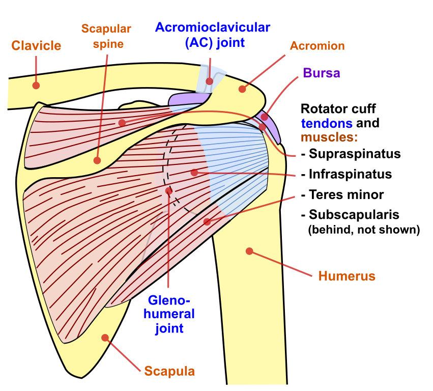
Diagram Of Shoulder Muscles And Tendons - Human Anatomy Body from www.anatomylibrary99.com 8 name the arteries and the nerves that supply shoulder joint. The shoulder is one of the largest and most complex joints in the body. There are five major shoulder ligaments that keep the shoulder in place and prevent it from dislocating. Webmd's shoulder anatomy page provides an image of the parts of the shoulder and describes its function, shoulder problems, and more. Ac joint is a diathrodial joint with a fibrocartilaginous disk. Although three ligaments protect and surround the shoulder joint, most of its stability comes from the powerful muscles and tendons of the rotator cuff. Notice superior labrum and attachment of the superior glenohumeral ligament. Shoulder muscles anatomy diagram see more about shoulder muscles anatomy diagram shoulder muscle diagram anterior shoulder muscles anatomy diagram muscles ligaments and tendons of the human back shoulder.
The transverse humeral ligament is not shown on this diagram.
Although the joint is held together by these extensive ligament and muscle attachments, certain types of forces can weaken the shoulder easily. Dr.g bhanu prakash animated medical. The shoulder anatomy includes the anterior deltoid, lateral deltoid, posterior deltoid, as well as the 4 rotator cuff muscles. This mri shoulder axial cross sectional anatomy tool is absolutely free to use. Webmd's shoulder anatomy page provides an image of the parts of the shoulder and describes its function, shoulder problems, and more. The shoulder is not a single joint, but a complex arrangement of bones, ligaments, muscles, and tendons that is better called the shoulder girdle. Robin smithuis and henk jan van der woude. The disk has a great variation in size and shape. The shoulder joint is formed where the humerus (upper arm bone) fits into the scapula. Shoulder bones and ligaments anatomy. Use the mouse scroll wheel to move the images up and down alternatively use the tiny arrows (>>) on both side of the image to move the images. There are many shoulder ligaments which each play an important role in shoulder joint stabilization to various degrees: Superior, middle and inferior ligaments, connect the glenoid to the anatomical neck of the humerus an.
Shoulder bones and ligaments anatomy. Normal anatomy, variants and checklist. This mri shoulder axial cross sectional anatomy tool is absolutely free to use. Webmd's shoulder anatomy page provides an image of the parts of the shoulder and describes its function, shoulder problems, and more. Roots, trunks, divisions, cords, branches, clinical anatomy.
File:Shoulder joint back-en.svg - Wikimedia Commons from upload.wikimedia.org The clavicle (collarbone), the scapula (shoulder blade), and the humerus (upper arm bone) as well as associated muscles, ligaments and tendons. One or more ligaments provide stability to a joint during rest and movement. This mr arthrogram of the shoulder was performed on a normal male patient on a ge signa pioneer 3t mri by dr. Choose from a wide range of similar scenes. You can see it enclosing the glenohumeral joint and you can see its attachment on the anatomical you've got the transverse humeral ligament and the coracohumeral ligament. Use the mouse scroll wheel to move the images up and down alternatively use the tiny arrows (>>) on both side of the image to move the images. Corey chakarun from shin imaging in california. Bones in shoulder, ligaments of the shoulder joint, parts of the shoulder joint, shoulder anatomy, shoulder joints and muscles, shoulder structure anatomy, shoulder tendon anatomy, shoulder related posts of diagram of shoulder muscles and tendons.
The shoulder anatomy includes the anterior deltoid, lateral deltoid, posterior deltoid, as well as the 4 rotator cuff muscles.
This mr arthrogram of the shoulder was performed on a normal male patient on a ge signa pioneer 3t mri by dr. Last update february 25, 2021. Additional stability is provided by: Notice superior labrum and attachment of the superior glenohumeral ligament. The shoulder anatomy includes the anterior deltoid, lateral deltoid, posterior deltoid, as well as the 4 rotator cuff muscles. Home > blog > anatomy > shoulder anatomy: A joint capsule is a watertight sac that surrounds a joint. Ligaments are fibrous bands or sheets of connective tissue linking two or more bones, cartilages, or structures together. Shoulder bones and ligaments anatomy. All about the shoulder muscles. The shoulder joint is formed where the humerus (upper arm bone) fits into the scapula. Ac joint is a diathrodial joint with a fibrocartilaginous disk. Human anatomy human body anatomy shoulder anatomy medical illustration human massage therapy ligament tear body anatomy for artists.
This mri shoulder axial cross sectional anatomy tool is absolutely free to use shoulder anatomy diagram. It is the major joint connecting the upper limb to the trunk.


0 Komentar